
This reference uses the specific documents linked in the article and listed below. Check their scope, publication dates and the statements they support.
The hoof is losing a supporting connection
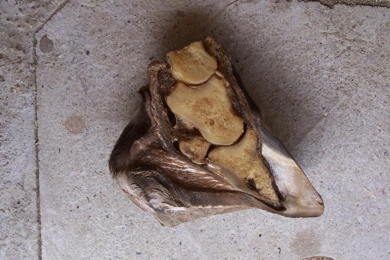
The Merck laminitis reference describes injury to the interlocking laminae that suspend the bony column within the hoof. When attachment weakens severely, the pedal bone can rotate or sink. Laminitis is therefore not simply heat in the foot or an overlong toe.
The distinction explains why the external appearance may understate an early problem. A horse can need urgent assessment before the hoof wall develops the changes associated with chronic disease. Preserve the onset of pain and the preceding illness or management history rather than dating the case only from the first radiograph.
Several pathways can converge on the same tissue
The American College of Veterinary Surgeons discusses systemic illness, dietary and metabolic circumstances, and excess loading of a supporting limb among the risk contexts. These are different pathways; finding laminitis does not by itself establish which one occurred.
A horse with endocrine risk, a mare unwell after foaling and a horse overloading one limb because the other is injured need different investigations. Keep the primary problem and the hoof complication linked without treating them as interchangeable diagnoses. This is also why a single pasture rule cannot prevent every form of laminitis.
Recognize pain without demanding a demonstration
Lameness, reluctance to move, an altered stance and increased digital pulse are among the clinical findings described by ACVS. None should be tested by forcing the horse through repeated turns or exercise. Ask the veterinarian how the animal should be kept comfortable and assessed safely.
Describe whether the difficulty appeared suddenly, involves several feet or followed another problem. A video of naturally occurring movement may help preserve an observation, but it should not require making an already painful horse walk for the camera.
Imaging answers a structural question
Merck explains how radiographs help assess the relationship of the pedal bone to the hoof capsule, especially in more established disease. Early and later images can show different information. A lack of major displacement on the first examination does not mean the painful process should be ignored.
Keep the original images, dates and veterinary interpretation with the farriery plan. A measurement copied from one image should not be treated as directly comparable with another taken differently unless the professional team considers the comparison valid. Comfort, structure and function are related outcomes, but they are not identical measurements.
Treat the cause and support the foot
Merck describes managing the underlying disease, pain, movement and hoof support within treatment. ACVS emphasizes that intervention can be most influential before extensive damage develops. The actual approach needs the veterinarian and appropriately qualified farrier working from the same assessment.
Do not improvise a trimming, shoeing or medication plan from a photograph or another horse's recovery story. The affected feet, structural findings and systemic circumstances can differ. Obtain clear instructions about movement, footing, feeding and reassessment, and preserve who is responsible for each part.
Recovery is a course, not one reassuring day
An illustrative horse may become more willing to stand after pain management while still needing structural monitoring and a plan for the initiating condition. Improvement is meaningful, but it should not be recorded as proof that all hoof damage has resolved or that exercise can resume without advice.
Keep changes in comfort, hoof assessment and work status separately. If several professionals are involved, maintain one shared sequence of decisions and the actual changes made. A missing communication about the latest image can undermine an otherwise carefully coordinated plan.
Questions about laminitis
Does every case come from rich grass?
No. Dietary and endocrine circumstances are important, but systemic illness and supporting-limb overload are other recognized contexts.
Must the pedal bone already be displaced?
No. Laminar injury and pain can precede major displacement. The examination and imaging determine the current findings.
Can a warm hoof diagnose it?
No. Warmth is nonspecific. Prompt veterinary assessment is appropriate when pain or the history raises concern. The horse hub and veterinary resources page provide connected reading and handoff support.
Cite this reference
LivestockDefense. “Equine laminitis: hoof support failure and urgent assessment.” Updated 2026-10-28. https://livestockdefense.com/threats/equine-laminitis/
Add the date you accessed the page. This identifies the current reference; keep a permitted saved copy if your work depends on its exact wording. A citation alone does not establish support for a claim.
How to cite a page and its revision ↗Sources & boundaries
Read the evidence in context.
These documents support the statements identified in this reference. Check their scope and dates when deciding whether they answer your question.
- Merck Veterinary Manual: Laminitis in Horses
Source update: 2024-06. Most recent check: 2026-10-28.
Laminar attachment injury, phases, radiography and treatment principles; no owner-directed trimming or medication plan.
- American College of Veterinary Surgeons: Laminitis in Horses
Source update: Not provided. Most recent check: 2026-10-28.
Risk pathways, clinical and radiographic assessment, early intervention and team-based support; no universal prognosis.