
This reference uses the specific documents linked in the article and listed below. Check their scope, publication dates and the statements they support.
External lumps and internal disease are two presentations
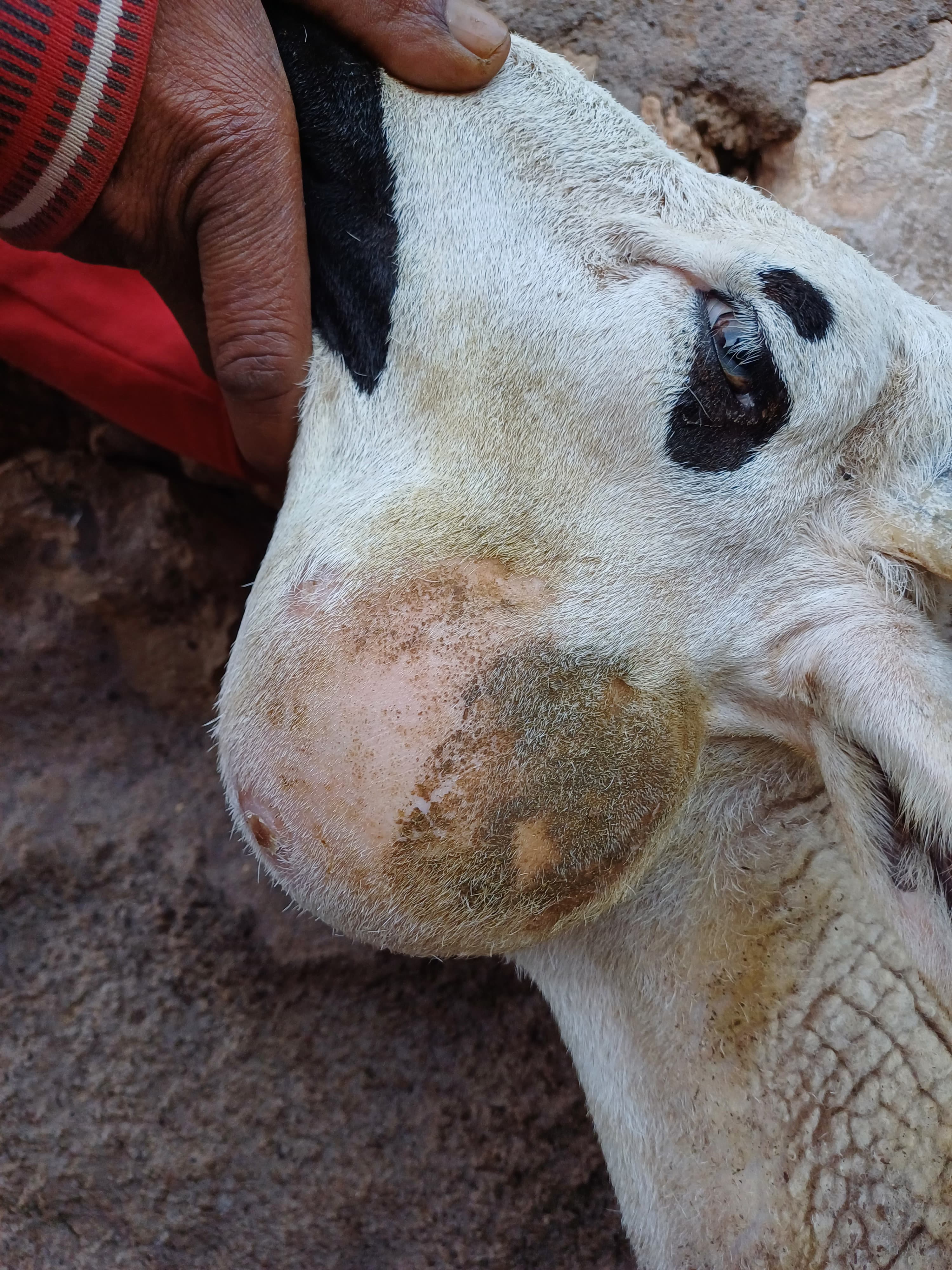
Peripheral lymph-node abscesses may appear around the head, neck or the origins of the limbs. Internal abscesses are harder to see and can affect organs or deeper lymph nodes. Both forms occur in sheep and goats; visible external disease is especially familiar in goats, while internal disease is an important concern in thin sheep. External and internal caseous lymphadenitis.
A lump is not automatically CL. Other bacteria, a local injury and other lesions can resemble an abscess. Similarly, a sheep losing weight despite access to feed may have Johne’s disease, gastrointestinal parasites, poor teeth or another chronic illness. The examination needs to explain the actual animal rather than rely on the herd’s previous diagnosis.
Internal infection can be present without a visible lump. This limits what a quick visual inspection of a purchase can establish. It also explains why resolving one obvious abscess does not prove that the animal has no deeper disease.
Why a draining abscess changes the flock’s exposure
Discharge can contaminate accommodation, equipment and surfaces used by other sheep or goats. Skin damage during shearing or other husbandry creates opportunities for entry. Animals with internal respiratory involvement can also contribute to exposure. Transmission and husbandry risk factors.
Separate a suspect animal before the lump ruptures when possible, and arrange assessment. Do not squeeze or lance a lump in a shared pen. Apart from the welfare and procedural risks, opening it can spread material onto the very surfaces that healthy animals subsequently use.
Review the shearing and handling sequence with the veterinarian and contractor. Equipment hygiene and avoiding accidental cutting into an abscess are practical priorities. A control plan that ignores how animals share tools and facilities is unlikely to succeed merely because visible cases are occasionally treated.
Diagnosis starts with confirming the organism
The veterinarian can arrange appropriate investigation of an active lesion to identify its cause. Laboratory identification of C. pseudotuberculosis provides stronger evidence than the position or appearance of the lump alone. Keep the animal separate while the cause is being established rather than waiting for a draining lesion to look “typical.” Diagnostic laboratory overview.
Blood antibody testing can contribute to a flock assessment, but individual interpretation is not absolute. Prior exposure, vaccination, age and maternal antibodies can affect results; false-negative results can also occur. A positive result is not a simple count of internal abscesses, and a negative result does not give an unconditional guarantee of freedom from infection. Serology limitations and clinical interpretation.
Internal disease may require imaging or further investigation. Discuss what each proposed test can answer and how the result changes management. Repeatedly testing without a decision about separation or breeding can leave the important exposure problem unchanged.
Treating a lesion is different from curing chronic infection
Treatment is difficult because abscesses and internal infection can persist. Veterinary options may include management of an accessible lesion, surgery in selected animals or an antimicrobial plan. These interventions do not reliably guarantee that infection has been eliminated or that no new abscess will appear. Veterinary abscess-management context; Noncurative treatment limitations.
Whether treatment is worthwhile depends on the animal’s welfare, lesion location, internal disease suspicion, the ability to maintain separation and the flock’s health objective. An individual breeding animal and a commercial flock pursuing freedom from CL may require different decisions, but neither should be represented as cured merely because the surface wound closes.
Do not inject formalin or other caustic preparations into a lump, and do not attempt surgery around major vessels and nerves. Food-animal drug authorization and withdrawal requirements remain relevant to any veterinary treatment.
A clear flock objective makes control more practical
Where the objective is to maintain freedom from CL, purchase from well-characterized sources, assess animals before entry and use a testing strategy appropriate to the source population. A seller’s statement that no abscess is visible is not equivalent to a supported flock-health status.
Where infection is already established, discuss separation or removal of infected animals, management of suspect lumps and reduction of environmental contamination. Internal cases and recurring disease can make control slow. Culling may be part of a program, but a single uncertain lump should first be evaluated rather than automatically assigned the diagnosis. Control principles.
Remove sharp environmental hazards and manage other causes of skin injury. Protect uninfected groups during shearing and handling. Shared sheep and goats need to be considered together rather than treating the species boundary as a barrier to infection.
Vaccination helps some programs but does not certify freedom
Suitable vaccines may reduce disease in particular settings, but availability and authorization vary by species and country. A sheep product is not automatically appropriate for goats. Vaccination can complicate antibody interpretation, so the veterinarian should coordinate the product decision with the testing and sourcing plan.
No vaccine should be promised to cure an infected animal or remove the need to control draining lesions. The useful question is how it fits a defined flock goal and what clinical benefit can realistically be expected.
Questions about handling and prognosis
Can people become infected?
Human infection is uncommon but possible, particularly with occupational exposure. Wear gloves and protect skin breaks when handling suspect animals or contaminated material; use pasteurized milk for human consumption. Seek medical advice for an unusual persistent lesion and mention the exposure. Public-health significance.
Can a sheep with no external abscess have CL?
Yes. Internal disease may cause chronic weight loss or respiratory illness. The sheep and goat hubs link to related chronic-disease differentials and husbandry guidance.
Cite this reference
LivestockDefense. “Caseous lymphadenitis in sheep and goats: abscesses, testing and flock control.” Updated 2026-11-02. https://livestockdefense.com/threats/caseous-lymphadenitis/
Add the date you accessed the page. This identifies the current reference; keep a permitted saved copy if your work depends on its exact wording. A citation alone does not establish support for a claim.
How to cite a page and its revision ↗Sources & boundaries
Read the evidence in context.
These documents support the statements identified in this reference. Check their scope and dates when deciding whether they answer your question.
- Merck Veterinary Manual: Caseous lymphadenitis of sheep and goats
Source update: 2024-09. Most recent check: 2026-11-02.
External/internal abscesses, differential and test interpretation, treatment difficulty and environmental control. No abscess opening, culture, chemical injection or invasive procedure reproduced.
- Louisiana State University AgCenter: Caseous lymphadenitis
Source update: Not provided. Most recent check: 2026-11-02.
Internal versus peripheral disease, shearing/contact exposure, noncurative treatment and control planning; no home drainage, disposal recipe or historical dip-additive suggestions reproduced.
- University of Minnesota: Abscesses related to systemic infections
Source update: Not provided. Most recent check: 2026-11-02.
Caseous lymphadenitis section: chronic infection and veterinary abscess/surgical judgment. Technical surgical content and antibiotic recipes are not reproduced.
- Washington State University Animal Disease Diagnostic Laboratory: Caseous lymphadenitis of sheep and goats
Source update: 2022-11-10. Most recent check: 2026-11-02.
Primary search-indexed diagnostic overview inspected; direct fetch returned 403. Limited to existence and clinical interpretation of agent identification and antibody testing, not unexposed procedural details.